Case Report - (2022) Volume 11, Issue 3
Perforation of one or more intraperitoneal organs during a liposuction procedure is an unusual and underestimated complication. Awareness of this complication is essential due to the frequent delay in diagnosis and potentially fatal consequences.We analyze the case of a 49-year-old woman who presented to the emergency room with septic shock of abdominal wall and necrotizing fasciitis developed after cosmetic surgery. The patient was admitted with a Glasgow score of 15 and generalized pallor, and was treated in the operating room for revision and cavity cleansing. Abundant intestinal contents were found in the subcutaneous cellular tissue, necrosis and liquefaction of Treitz tissue up to 20 cm of the ileocecal valve, peripancreatic edema and multiple wheals in the omentum. On the 42nd day of hospital stay, it was decided to perform fistula management by the Rivera condom technique and graft placement. Among the pathogens identified in culture, the most important are E. Coli, A. Baumanni multidrogoresitente, Candida famata, which are managed favorably. The patient was discharged on the 77th day of her hospital stay.Visceral injury, although extremely infrequent, is a real possibility following liposuction. Post-surgical follow-up should be cautious, with surveillance of abdominal or thoracic symptomatology. Its dramatic appearance and the frequent delay in diagnosis make it essential to be aware of this complication.
Liposuction • Visceral perforation • Complications • Rivera condom • Stoma management
In the five countries that perform the highest number of aesthetic surgical procedures (United States, Brazil, China, Japan, and Mexico), liposuction is the most frequent [1]. Nowadays, the literature reports a low incidence of both local and systemic complications of liposuction, among the latter the most serious are: deep vein thrombosis, pulmonary embolism, cavity perforation, necrotizing fasciitis, sepsis, and infarction [2]. Penetration of the abdominal wall and consequent injury of one or more viscera is rare and underestimated, but represents a life-threatening complication. Knowledge of this type of complication is essential to treat the patient quickly and avoid dramatic consequences [3].
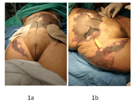
The observations corresponding to this case took place at the “Regional Hospital No. 20 of the Mexican Social Security Institute”, all data were extracted with due authorization and are stored in the hospital's records. Autograph consent was obtained from the patient for the presentation and publication of her case. We present the case of a 49-year-old female patient, native of the city of Zapopan, Jalisco, who was admitted to the intensive care unit with the following diagnoses: abdominal septic shock, post-operated drainage plus surgical wound cleansing and debridement, colon raffia with sigmoid and epiploic appendix patch in open abdomen status. Four days before her admission to our service, the patient underwent laparotomy, due to perforation of hollow viscera from a liposuction procedure. The patient was not a carrier of chronic degenerative diseases. She had undergone lipectomy and mastopexy with abdominoplasty and lipo-transfer 12 years ago. She only mentioned that she was allergic to ibuprofen. The patient underwent a cosmetic procedure (liposuction and lipectomy), in which complications of perforation of the hollow viscera occurred. Considering this situation, a laparotomy was performed where the following findings were found: abundant free fluid in a cavity with intestinal characteristics, multiple perforations from the ligament of Treitz to 10 cm of the ileojejunal vulva. A 100 cm of jejuno-ileum was dissected and later a laterolateral anastomosis was performed with a linear stapler. Finally, the anastomosis mouth was closed with cavity lavage, using 8 liters of solution, and Penrose was placed. The patient was transferred to a private medical unit for intensive therapy management where she was administered crystalloid solutions, vasopressors, supplementary oxygen support, piperacillin, tazobactam, linezolid, and fluconazole. On the fourth day of her current condition, the patient was admitted to our emergency department, with a Glasgow score of 15 points, neurologically integrated, with normal blood pressure, but generalized pallor. At that moment, the general surgery service decided to manage her in the operating room, finding abundant intestinal content in the subcutaneous cellular tissue, necrosis and liquefaction of small intestinal loops from 20 cm from the fixed loop (treitz angle) to 10 cm before the ileocecal valve, peripancreatic edema, multiple bruises in the omentum; anesthesiology reported mild bleeding without the need for hematotransfusion (Figures 1a and 1b).

Figure 1: 1a) Patient is received in the emergency room with abdominal septic shock 1b) Necrotizing fasciitis of the abdominal wall is observed.
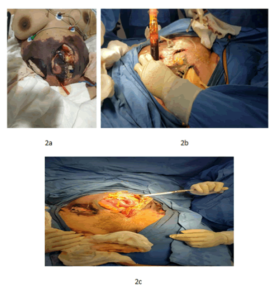
The patient was admitted to the ICU with priority IA; it was decided to deepen the sedation and an orogastric tube was placed for a biliary content derivation; an open abdomen was observed, with an open LAPE type surgical wound, with compresses inside, as well as drainage in the lower part of the wound, ecchymosis in the entire abdominal region and a second surgical wound in the right iliac fossa of approximately 15 cm. One day later, sedation was maintained, and the diagnosis from the Intensive Care Unit department was severe sepsis with abdominal septic shock, bowel perforation, necrotizing fasciitis, and acute renal failure, with a poor short-term vital prognosis. The patient was on her eighth day of in-hospital stay, in the intensive care unit, with the following diagnoses: Abdominal departure septic shock, Post-Operative (PO) tracheostomy/gastrostomy/ surgical lavage, necrotizing fasciitis of the abdominal wall, PO surgical lavage with debridement, PO surgical wound abscess drainage with sigmoid colon raffia, PO exploratory laparotomy, acute kidney injury, metabolic acidosis with respiratory alkalosis and moderate normocytic normochromic normocytic anemia. On the10th day, a Mahurkar catheter was placed to start the hemodialysis session. On the 13th day, surgical cleaning was performed with placement of a negative pressure therapy system, the wound remained open at the end of the procedure (Figures 2a-2c); On the 17th day of HIE, infection was found in the surgical wound, it was scheduled for drainage, cleaning, and debridement of the wound and replacement of negative pressure therapy.

Figure 2: 2a) Patient undergoing his first check-up and cavity cleaning 2b) First admission to the operating room for review and surgical cleaning 2c) Surgical cleaning with placement of negative pressure therapy system.
Surgical technique
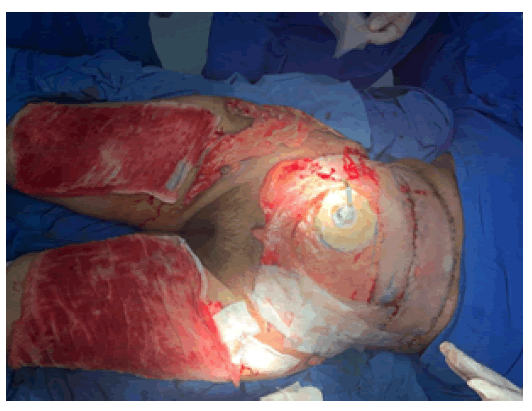
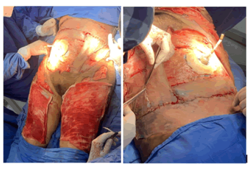
Under sedation, previous asepsis, and antisepsis, grafts were taken from the right leg, which was placed in the abdomen over the cruciate region (in abdominal and inguinal regions), fixed with 3-0 nylon with simple stitches. A Rivera condom was placed in the stoma and the colostomy bag was left free (Figures 3 and 4). The grafts placed showed a good evolution.

Figure 3: Fistula management with Rivera's condom technique.

Figure 4: Placement of grafts.
Recently Gardener et al. presented a case report of a 69-year-old woman who underwent abdominal contouring surgery consisting of correction of a flank pseudohernia, liposuction, and short scar abdominoplasty, which was complicated by intestinal perforation [4]. They propose that the position of the patient on the operating table and the laxity of the abdominal wall during surgery, as well as the timing of each specific procedure, play an important role in the occurrence of bowel perforation. Pohlan et al. described a year later a case of multiple liver perforations in a patient who underwent an outpatient liposuction procedure, was treated without surgery and was discharged after 7 days. The author insists that, despite the infrequency of these complications, patients should be monitored especially if they present abdominal symptoms [5]. Risks for perforation of viscera during liposuction have been described to include morbid obesity, previous surgical scars, diastasis of the rectus abdominis, and abdominal wall hernias. Perforation may extend to surrounding lymphatic, vascular, and intra-abdominal structures, or may occur far from the original surgical site, as in the case of patients with severe chest pain and dyspnea, possibly indicating perforation [6].
On the other hand, postoperative flap involvement is usually due to insufficient tissue perfusion secondary to disruption of the subcutaneous perforator vessels and the subdermal plexus, as happened to our patient. This can lead to a variety of acute complications depending on the depth of tissue involvement. Epidermolysis is the mildest variant in which only the epidermis suffers ischemia [7]. The natural course of uncomplicated epidermolysis is spontaneous re-epithelialization without intervention, however, more severe necrosis can lead to tissue loss and the need to use other strategies, as in the case we present, where we rely on graft placement. Regarding the management of our patient's stomas, in patients with hostile abdomen, intestinal perforations are frequent and are managed by primary closure and/or splinting with probes; however, leakage of intestinal material is not controlled in all cases. However, at the end of the 19th century, peritonitis was treated medically with a mortality rate of 90%; in 1926, Krishner demonstrated that this could be reduced with the strict implementation of surgical principles, and the mortality rate fell below 50% [8]. It is during the process of resuscitation and stabilization of the patient where the method of externalization of the fistula using a latex tube (placement of a condom) is efficient since it helps to control the septic focus by preventing the leakage of intestinal contents into the abdominal cavity, which reduces peritoneal irritation and, therefore, the systemic inflammatory response [9]
It is a fact that aesthetic procedures are especially lucrative. This is a situation that we must consider because lacking legal restrictions, they are increasingly performed in outpatient settings, by non-plastic and even nonphysician surgeons [10]. Growing medical tourism has stimulated demand for cosmetic surgery in Europe, South America, and Southeast Asia. A survey distributed to 2000 active members of the American Society of Plastic Surgeons (ASPS) showed that 51.6% of respondents noted an increasing trend in the number of patients presenting with complications from surgical tourism [11]. The case we present was originally attended by a physician not specialized in plastic and aesthetic surgery, who performed this second liposuction. At present, there is no accurate data on how many of these procedures are performed by non-plastic physicians, which may leave a wide margin lacking supervision and, therefore, a risk of morbidity and mortality. It is common that, in these cases, the physician does not evaluate these patients until three or four days after surgery; therefore, they usually go to the emergency department with advanced complications [12].
Perforation of one or more intraperitoneal organs during liposuction is relatively rare, but is underestimated and is probably the most serious of all. Its dramatic appearance and the frequent delay in diagnosis make it essential to be aware of this complication. It is proposed to maintain current statistics involving the reporting of post-surgical complications of elective procedures, supervision of known centers in compliance with good clinical practices in order to avoid this type of complications derived from negligence.
Competing Interests
The authors declare that they did not receive any type of financing from any institution. They also declare that there is no conflict of interest for the publication of this clinical case.
The authors declare that they have obtained the consent of the patient for the publication of this case and it is made available if the editor so requests. All data and images obtained here have sought to safeguard the integrity and confidentiality of the patient.
Citation: Carbajal, S., J. et. al. Visceral Perforation Following Liposuction and Management of Hostile Abdomen with the Rivera Condom Technique. Case Report. Reconstr Surg Anaplastol. 2022, 11(3), 001-003.
Received: 28-Jun-2022, Manuscript No. ACR-22-19005; Editor assigned: 30-Jun-2022, Pre QC No. ACR-22-19005 (PQ); Reviewed: 07-Jul-2022, QC No. ACR-22-19005 (Q); Revised: 15-Jul-2022, Manuscript No. ACR-22-19005 (R); Published: 28-Jul-2022, DOI: doi.10.37532/22.11.3.1-3
Copyright: ©2022 Carbajal, S., J. This is an open-access article distributed under the terms of the Creative Commons Attribution License CC-BY, which permits unrestricted use, distribution, and reproduction in any medium, provided the original author and source are credited.