Case Report - (2023) Volume 12, Issue 2
PORPOSE: Reconstruction of the anatomical area of the knee is challenging due to its intrinsic needs in terms of vascularization, reliability an pliability of the covering tissues. In a difficult setting where the classic workhorses flaps have failed to cover the defect a new approach is needed to overcome the limits of well known salvage procedures.
CASE PRESENTATION: A 16 years old girl underwent surgery for a tibial head osteosarcoma. Classical local flaps failed to cover the defect in other institutions due to different problems and the the prothesis was substituted by antibiotic-loaded bone cement. The defect measured 5x4 cm. The amputation was avoided by using the vastsus intermedius flap.
DISCUSSION: At 5-day post-op, neither necrosis of the flap nor dehiscence of the wound was detected. After two months the definitive prothesis was inserted. At two-years follow up the flap tissue is soft and pliable, the coverage is optimal and the patient can walk without crutches. In a difficult setting the vastus intermedius flap is reliable solution useful to spare the few remaining vessels of the area for a future salvage procedure. This research did not receive any specific grant from funding agencies in the public, commercial, or not-for-profit sectors.
Compliance with Ethical Standards
Conflict of Interest: The authors declare not to have any conflict of interest. Informed consent was obtained from the individual included in the report.
Plastic Surgery • Osteosarcoma • Oncological Surgery • Vastus Intermedius Flap
The anatomical area of the knee is a major challenge for the reconstructive surgeon due to its structural and functional complexity [1]. Thin, pliable and well vascularized tissues are required for covering the joint or a prosthesis [2, 3].
Furthermore in the oncological setting a multiple stage surgery with a focus concerning donor site morbidity is often mandatory due to the need of radicalization or high recurrence rate. The surgeon has always to bear in mind the reconstructive ladder and should consider different surgical approaches not to waste the tissue required for a subsequent step in the surgical treatment. Chemotherapy or RT additional therapies should also taken into account by reason of their detrimental effect on wound healing.
The authors propose a new muscular flap for replacing soft tissue defects of the knee. This solution was employed for the first time for covering a antibiotic-loaded bone cement area of the knee in a young patient affected by osteosarcoma suffering prosthesis exposition after both bilateral gastrocnemius flap and local flaps failure. The decision to use this innovative flap was taken in order to spare a microsurgical flap for an eventual coverage of the amputation stump.
A 16 years old girl underwent surgery for a tibial head osteosarcoma in another Hospital. A knee prosthesis was inserted and primary closure was performed.
After the exposition of the prosthesis a lateral gastrocnemius flap was attempted but failed for sufferance of the distal third and then a medial gastrocnemius flap was also spent to cover the medial part of the residual defect but partly failed due to infectious complications.
A prosthesis substitution with antibiotic-loaded bone cement was made after leg shortening. The soft tissue defect was closed primarily.
The patient finally came to our Hospital, where exposition of the antibiotic-loaded bone cement with a defect of 5 cm × 4 cm (Figure 1) led the orthopedic surgeon to consult our Team in order to perform a combined amputation and flap coverage of the stump.
Figure 1: Preoperative aspect
We initially decided to try a salvage coverage with a free flap based on a reliable recipient vessel found in the area of the defect. Considering the high risk of amputation of the limb in a further operation we figured out the possibility of using a viable flap found during the dissection in order to save the donor site area.
Written informed consent was achieved and the the procedure started as stated above.
During the search of recipient vessels a trophic vastus intermedius muscle unnecessary for the stability of the immobilized inferior limb was found.
We decide to use the vastus intermedius flap to cover the defect. The anatomy of vastus intermedius is shown in cadaveric study in Figure 2.
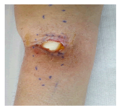
Figure 2: Vastus intermedius muscle, cadaveric dissection
The flap completely healed and the stitches were removed after 21 days. The patient was discharged in the fifth day post-op and came to the outpatient department once a week for medical dressing. Cephazoline therapy at 2 g per day was administered for 14 days. After 2 months the previous surgical access was used to allow the orthopedic surgeon to substitute the antibiotic-loaded bone cement with a definitive prosthesis. During a two-years follow up examination the flap is still good, the tissue is soft and pliable, the coverage is optimal (Figures 3,4) Furthermore the patient walks without crutches.
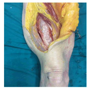
Figure 3: Follow-up at two years frontal aspect.
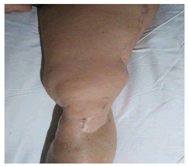
Figure 4: Follow- up at two years later aspect.
The reliability of this muscle as a flap was investigated with a handheld Doppler probe and then it was dissected tracing its multiple nourishing vessels originated from the lateral circumflex femoral artery. The dissection proceeded in a proximal to distal way and after the pedicles were exposed progressively the vessels were clamped starting from the most cranial to the caudal one, collocated a few centimeter from the rotula.
During the clamping maneuvers a vitality test of the connected area of the muscle was performed by waiting some minute and scaring the surface of the muscle. The corresponding pedicle is then ligated and the vastus intermedius was detached from the femoris.
The flap is turned 180° arch on its longitudinal axis leaving attached the two most caudal vessels and the defect was comfortably covered after a proper surgical debridement. (Sequence is shown in Figures 5-7).
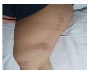
Figure 5: Vastus medialis vastus intermedius and vastus lateralis in site
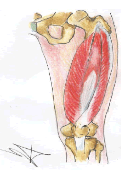
Figure 6: Vastus intermedius arc of rotation around its pivot
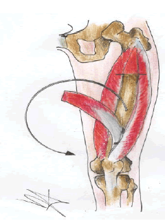
Figure 7: Vastus intermedius after complete rotation for defect covering
The survival of the flap was checked once again, hemostasis was performed and then the flap was covered with a split thickness skin graft and sutured with a tie-over dressing. The muscle margins were inserted beyond the skin edges of the debrided area. No drains were positioned. Cephazoline was administered as prophylactic therapy.
The limb was blocked with a valve for three weeks and the patient was no weight bearing was allowed during that period.
Oncological resection represent one of the most frequent causes of soft tissue defects in the knee region.
The need for wide resection or multiple surgical procedures often lead to a total arthroplasty with wound a complication rate up to 20% with soft tissue tissue sufferance and implant exposure. In some cases, the development of a severe infection may lead to prosthesis loss or amputation [4-6]. Approaching knee surgery is important to bear in mind the general condition of the patient (e.g infection and dehiscence related to diabetes and deep venous thrombosis related to obesity) [6-8].
In fact oncologic patients are often affected by comorbidities, a poor general status due to multiple treatments and negative local factors such as previous scars, damaged vessels and irradiated soft tissues.Furthermore aggressive cancers are burdened with a high recurrence rate that demands another subsequent surgical reconstruction. Unfortunately amputation is a common scenario and a stump coverage with a reliable solution is demanded. Recent studies demonstrate that is preferable to try a salvage surgery as it has higher 5-year survival rates, while not increasing the risk of local recurrence [9].
An anatomically and functionally complex region like this requires strong and pliable tissue to replace the like with like, minimize donor-site morbidity preserve main vascular trunks, and reduce operating and hospitalization time [10].
Nevertheless in complex demolitions is sometimes impossible to find a viable standard local flap and the reconstructive surgeon is forced to use a microvascular flap. Free flaps are challenging procedure in the anterior knee area especially in case of an impaired perfusion limb. Furthermore a sound expertise and proper tools are needed [11], and besides is better to spare a free flap for a salvage procedure. Another aspect that must be evaluated before choosing a free flap as first option in this previously operated region is the recipient vessel issue: a correct vessel selection is the cornerstone of success of a free flap [12]. In a inferior limb damaged by multiple surgeries, therapies, infections and surgical toilettes a good vessel selection is a challenging point. It could be impossible to find a reliable one or during the procedure and there is a high risk to compromise the few survival vessels that could sustain a salvage procedure. The vastus intermedius flap perfectly obliterated the defect, introduced a well-perfused tissue from an uninjured area, thus facilitating wound healing and antibiotics delivery.
It allowed us to overcome all the above mentioned problems and to obtain a solid coverage without complications with a fast and reliable procedure.
New datas and surgical applications of the technique are surely needed but we think that this new flap could enrich the reconstructive armamentarium in selected difficult cases that already undergone many different procedures.
Conflict of Interest
The authors declare not to have any conflict of interest. Informed consent was obtained from the individual included in the report
Received: 01-Jan-2023, Manuscript No. ACR-23-23837; Editor assigned: 03-Jan-2023, Pre QC No. ACR-23-23837 (PQ); Reviewed: 18-Jan-2023, QC No. ACR-23-23837 (Q); Revised: 21-Jan-2023, Manuscript No. ACR-23-23837 (R); Published: 30-Jan-2023, DOI: 10.37532/acr.23.12.1.001-003
Copyright: ©2023 Fracon S. This is an open-access article distributed under the terms of the Creative Commons Attribution License, which permits unrestricted use, distribution, and reproduction in any medium, provided the original author and source are credited.