Case Report - (2020) Volume 5, Issue 3
History: A nine year old school girl, medically fit, complained of rash over her left axillae for 10 days. The patient was in her usual state of health until she noticed a rash that started gradually over the left axillae as solitary annular eczematous lesion which started to disseminate to the ipsilateral hemi thorax, abdomen and left thigh following dermatome, not crossing the midline, associated with itchiness and pain especially after sweating. The patient had no history of contact with topical medication, deodorant or any antiperspirant, no history of same lesion before, no history of medication exposure, no history of animal contact, no family history of same lesion, no history of fever, throat pain, cough or runny nose and other systemic review was unremarkable. Examination: Patient looked well, afebrile, vital signs were all with in normal. Over the left axillae there was a solitary 4 × 5 cm annular erythematous to brownish eczematous lesion covered with fine scales over the periphery of the lesions, surrounded by other smaller erythematous papules and plaques covered with fine scales that goes down to the ipsilateral trunk and one solitary plaque 2 × 3 cm over her left thigh. The right axillae, trunk and thigh were unremarkable. Wood lamp test was done over all the lesions which came negative. Investigation: Laboratory results showed a complete blood count and chemistry within the normal ranges. Bacterial cultures and titters were negative as were also viral serology, including those for herpes simplex virus 1 and 2, parvovirus B19, Cytomegalovirus (CMV), human herpes virus 6 and 7, and Epstein-Barr Virus (EBV). Skin biopsy was suggested by the physician but the parents refused as they know about the disease process. Management: Parents were educated about the disease process, skincare routine for such area, and to avoid topical irritant if any. Topical corticosteroid with miconazole was applied twice a day for two weeks, Fucidin cream twice a day for 5 days, Antihistamine syrup once a day PRN. The patient did not come to the follow-up due to (COVID 19) pandemic at the time but her mother sent us the picture of the lesion through teledermatology services. The patient is improving by our approach and all lesions have started to resolve on its own.
Unilateral laterothoracic exanthema; Sars-CoV-2; Rash; Case report
Unilateral laterothoracic exanthem is an uncommon skin rash that occurs most commonly in children. It is characterized by unilateral exanthem, often affecting the axillary region. The cause is unknown but a viral agent is suspected [1]. We report a 9-year-old girl with unilateral laterothoracic exanthem that occurs during the coronavirus pandemic SARS-CoV-2.
The disease was first described in 1962 and the term “Unilateral Laterothoracic Exanthem” (ULTE) was introduced in 1992. It occurs most commonly in the age range of 6 months to 10 years with a male: female ratio around 1: 2. The cause of ULTE remains unknown although viral etiology has been suggested screening for EBV, CMV, HHV-6, and HHV-7 was performed and no etiologic agent has been consistently demonstrated. An infection with Spiroplasma, parvovirus B19, and EBV has been noted in individual cases. The seasonal pattern has been reported, and it is usually occurring in winter or springtime. Unilateral Laterothoracic Exanthem (ULTE) is characterized by localized, unilateral exanthem, usually affecting the axillary region, which spreads in a centrifugal pattern, sometimes involving the contralateral side [1]. Sometimes it may be preceded by a prodromal upper respiratory or gastrointestinal upset [1]. The mucous membrane, face, palms, and soles are usually not affected [2,3]. The duration of eruption persists for around 4 to 6 weeks, exceptionally more than 8 weeks. Mild local lymphadenopathy has been found in about 50% of cases [1]. A skin biopsy is usually not characteristic. The histopathologic examination of biopsy may show perivascular and peri appendageal lymphocytic infiltrate, mononuclear cell exocytosis, spongiosis, and lichenoid dermatitis with parakeratosis [1].
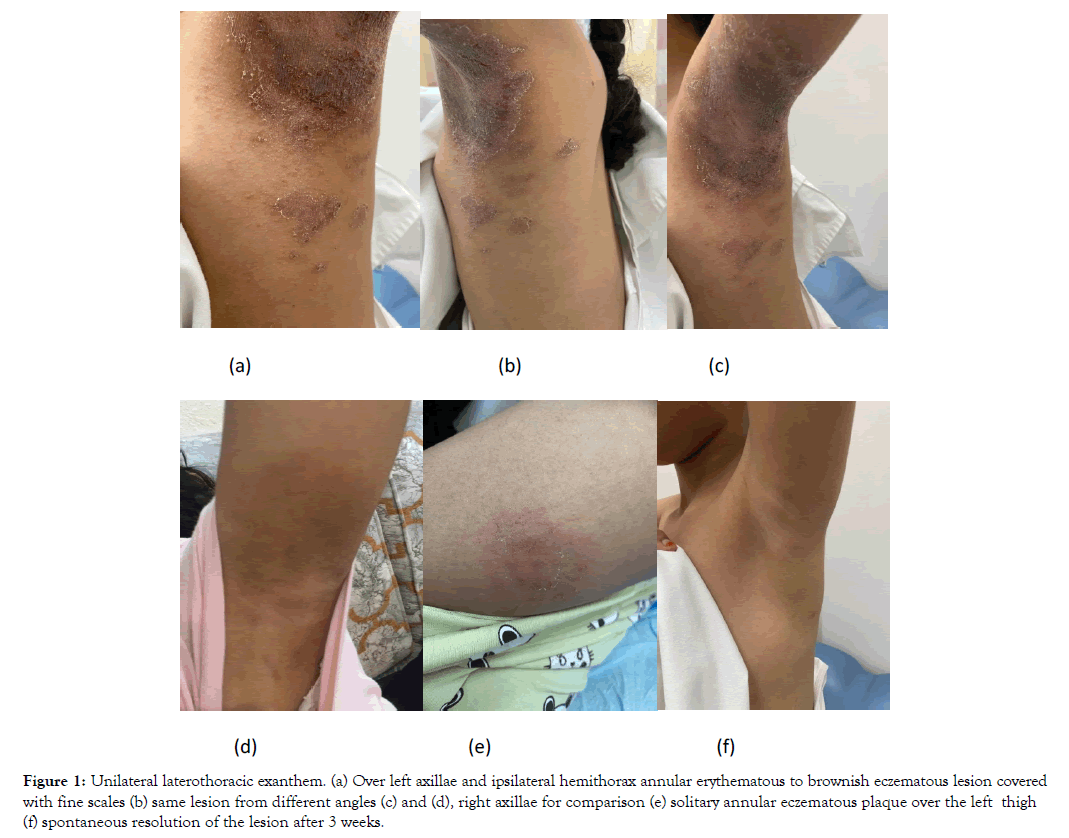
A 9 years old schoolgirl, medically free was referred to our Dermatology Department during the SARS-CoV-2 pandemic for an acute thoracic exanthema over her left axillae evolving for 10 days. The patient was in her usual state of health until she noticed a rash that started gradually over the left axillae as a solitary annular eczematous lesion that started to disseminate to the ipsilateral hemi thorax, abdomen, and left thigh following dermatome and not crossing the midline. It was associated with itchiness and pain especially after sweating. There was no history of applying any topical medication, deodorant, or antiperspirant, no history of animal contact or contact with a sick patient, no history of fever, throat pain, cough, or runny nose, and other systemic review was unremarkable. On examination, the patient looks well, afebrile, and vitally stable. Over the left axillae and ipsilateral hemi thorax, there is a 4 × 5 cm annular erythematous to brownish eczematous lesion covered with fine scales over the edge, surrounded by smaller coalescing erythematous papules and plaques. Over the left thigh, there is solitary annular eczematous plaque 2 × 3 cm with fine scales over the edge. The wood lamp test was negative. The remaining tegument examination was unremarkable and there was no mucous membrane involvement, the remaining physical examination, including lymph nodes was normal. The patient’s initial laboratory data showed a complete blood count and chemistry within the normal ranges bacterial cultures and titters were negative as were also viral serology, including those for herpes simplex virus 1 and 2, parvovirus B19, Cytomegalovirus (CMV), human herpesvirus 6 and 7, and Epstein-Barr Virus (EBV). A skin biopsy was suggested, but the parents refused when we explained the diagnosis and the nature of the disease to them. History, physical examination, and laboratory findings were consistent with the diagnosis of ULTE. We educate the parents about the disease process, skincare routine for such area, and avoid topical irritants if any. Antihistamine syrup once a day PRN, if there is itchiness, was given. She was started on topical corticosteroid with miconazole twice daily, Fucidin cream twice daily by the primary care physician before referral to us, and we suggest discontinuing those medications. The patient didn't come to the follow-up due to the SARS-CoV-2 pandemic at the time but her mother sent us the picture of the lesion through teledermatology services the patient was improving by our approach and all lesions started to resolve by itself (Figures 1a-1f).

Figure 1: Unilateral laterothoracic exanthem. (a) Over left axillae and ipsilateral hemithorax annular erythematous to brownish eczematous lesion covered with fine scales (b) same lesion from different angles (c) and (d), right axillae for comparison (e) solitary annular eczematous plaque over the left thigh (f) spontaneous resolution of the lesion after 3 weeks.
Unilateral laterothoracic exanthem is a self-limited disease that usually spontaneously resolves in about 3 to 6 weeks. UTLE may be preceded by nonspecific general symptoms, including fever, diarrhea, or rhinitis [4]. Most of the unilateral laterothoracic exanthem is seen in children, but it may affect adults also. Usually, it is characterized by a unilateral, axillary localized exanthem. Sometimes it may affect the contralateral side. The etiology of unilateral laterothoracic exanthem is unknown [5]. A possible relationship with Spiroplasma infection has been speculated by Taieb et al., [6] but it has not been confirmed. Other associations with bacterial, viral infections and vaccinations have been suggested but none of these associations could be proved. Some authors believe that the associations of unilateral laterothoracic exanthem with viral infections may be coincidental rather than having a causal relationship since viral infections are common in children. The seasonal pattern, early age of onset, associated prodrome, the lack of response to antibiotics, as well as the possibility of familial cases, suggest an infectious cause. Unilateral laterothoracic exanthem is a specific skin eruption that can be associated with parainfluenza 2 and 3, adenovirus, parvovirus B19, human herpes 6 and 7, and other microorganisms, such as and reactivation of EBV or CMV. Usually, no specific treatment is needed for unilateral laterothoracic exanthema as it will resolve spontaneously. Supportive treatment can be used for symptomatic patients although topical corticosteroids tend to be of little help [5-8].
In studies in which screening for multiple viruses was performed, no etiologic agent has been consistently demonstrated a relationship to infection with Spiroplasma, parvovirus B19, and EBV has been noted in individual cases like in our patient. However, more reports are needed to confirm this.
None
Citation: Almatrafi SF, Aljabri MM (2020) Unilateral Laterothoracic Exanthema: Case Report. Dermatol Case Rep 5:169.
Received: 06-Nov-2020 Published: 30-Nov-2020, DOI: 10.35248/2684-124X.20.5.169
Copyright: © 2020 SF Almatrafi, et al. This is an open-access article distributed under the terms of the Creative Commons Attribution License, which permits unrestricted use, distribution, and reproduction in any medium, provided the original author and source are credited.