Case Report - (2024) Volume 9, Issue 6
Sarcoidosis is a systemic disease of unknown origin affecting patients aged between 25 years and 40 years, and it has a higher incidence in women and in patients of African descent. We report the first case of sarcoidosis of the glans penis and penis, without systemic manifestations, in a patient of North African descent. Local treatment was initiated in this case. Regular monitoring should be performed to anticipate possible systematization.
• Sarcoidosis • Manifestations • Granulomatous disease • Penis • Traum
Sarcoidosis is a multisystem granulomatous disease with heterogeneous clinical manifestations. We report the case of a 46 year old man with sarcoidosis nodules of the glans penis and penis as the only manifestation of the disease [1].
A 46-year-old patient of North African origin presented with 5 asymptomatic infiltrated nodules of the glans penis, balanopreputial groove and penis for 8 months without any particular trauma. There were no other lesions. He was previously treated with antimycotic ointment for one month and then with ointment containing a low potency corticosteroid without any resolution; the nodules became more pronounced over time. The patient had a significant medical history of insulin independent diabetes that was well controlled by metformin for 5 years and epilepsy that was well controlled by carbamazepine and lamotrigine for 11 years [2].
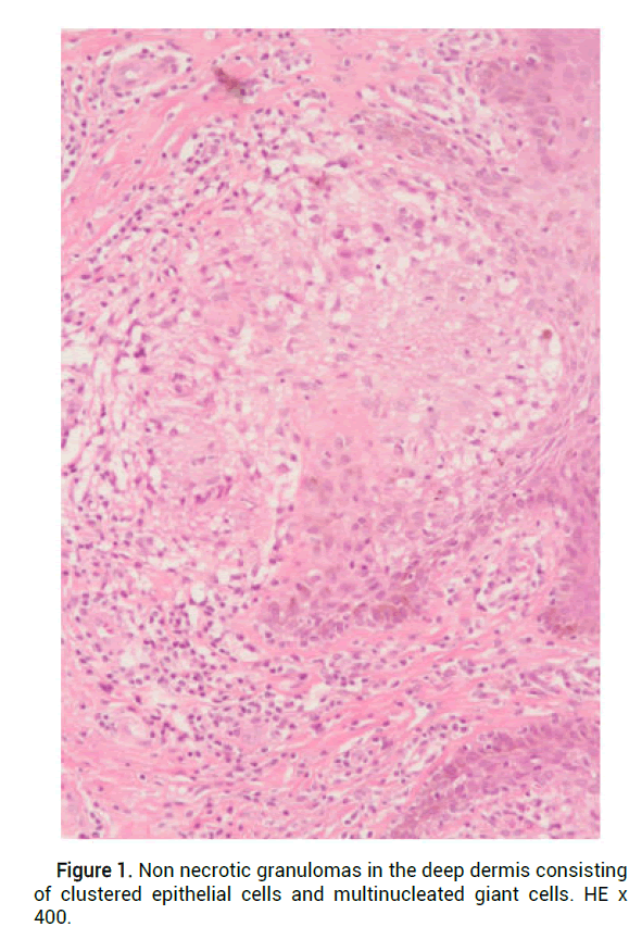
Histological examination of a biopsy of the lesions showed the presence of nonnecrotic granulomas in the deep dermis consisting of clustered epithelial cells and multinucleated giant cells. Tests for foreign bodies and specific infectious agents were negative (PAS, Gram, Zheil, Treponema).
The Mycobacterium culture was negative. A diagnosis of sarcoidosis was suggested. A chest X-ray was normal, as was a haematological examination. The Angiotensin Converting Hormone (ACTH) level was slightly above normal. An intradermal tuberculin test was normal. The diagnosis of nonsystemic sarcoidosis with a genital location was therefore maintained. An intralesional injection of triamcinolone acetonide 40 mg/ml cleared the disease without recurrence after 2 years [3].
Sarcoidosis is a systemic disease of unknown origin that affects patients aged between 25 and 40. A higher incidence is seen in women and in patients of African descent; African Americans often have more severe forms. Sarcoidosis can affect one or more organs and is characterized by the presence of noncaseating granulomas. Approximately 25% of patients with sarcoidosis have a cutaneous manifestation, while 90%-95% of patients have pulmonary manifestations [4]. The skin lesions of sarcoidosis have different clinical aspects: Macules, papules, plaques, and nodules. The occurrence of sarcoidosis on scars has been described, as well as after tattooing or after herpes zoster infection [5]. Indeed, any trauma to the skin can induce sarcoidosis in a predisposed patient. The presence of a single subungual sarcoidosis nodule with bone involvement has been described [6]. Genital involvement is rare; a penile mass and involvement of the epididymis, prostate, or testes have been described and are often seen in the later stages of the disease. However, scrotal and penile oedema has been described in nonsystemic sarcoidosis [7]. Sarcoidosis in the form of subcutaneous papules or nodules usually occurs in the earlier stages of systemic disease. There are 5 reports in the literature of penile sarcoidosis in the form of papules in the context of systemic sarcoidosis, which may also manifest as indurated oedema [8]. To our knowledge, this is the first case of sarcoidosis of the glans penis without systemic manifestations at the 2 years follow up (Figure 1). Due to the unique location, an injection of triamcinolone acetonide was performed with complete disappearance of the lesions [9-12].

Figure 1: Non necrotic granulomas in the deep dermis consisting of clustered epithelial cells and multinucleated giant cells. HE x 400.
We report the first case of sarcoidosis of the glans penis without systemic manifestations. Local treatment was initiated in this case. Regular monitoring should be performed to anticipate possible systematization.
Chloe Algoet (MD): Manuscript writing.
Olivier Vanhooteghem (MD): Project development, mauscript writing, manuscript editing.
Anne Theunis (MD): Data managment. Antoinette Henrijean (MD): Project development, manuscript writing.
N/A
N/A
Patient gives written informed consent to publish his data in this observational case report.
[Crossref] [Google Scholar] [PubMed]
[Crossref] [Google Scholar] [PubMed]
[Google Scholar] [PubMed]
[Google Scholar] [PubMed]
[Crossref] [Google Scholar] [PubMed]
[Crossref] [Google Scholar] [PubMed]
[Crossref] [Google Scholar] [PubMed]
[Crossref] [Google Scholar] [PubMed]
[Crossref] [Google Scholar] [PubMed]
[Crossref] [Google Scholar] [PubMed]
[Crossref] [Google Scholar] [PubMed]
[Crossref] [Google Scholar] [PubMed]
Citation: Algoet C, et al. "Nodules of the Glans Penis and Penis as the Only Manifestation of Nonsystemic Sarcoidosis; an Exceptional Location". Dermatol Case Rep, 2023, 8(5), 1-2.
Received: 08-Jul-2024, Manuscript No. DMCR-23-22167; Editor assigned: 10-Jul-2024, Pre QC No. DMCR-23-22167 (PQ); Reviewed: 12-Jul-2024, QC No. DMCR-23-22167; Revised: 16-Jul-2024, Manuscript No. DMCR-23-22167 (R); Published: 31-Jul-2024
Copyright: �© 2023 Algoet C, et al. This is an open-access article distributed under the terms of the Creative Commons Attribution License, which permits unrestricted use, distribution, and reproduction in any medium, provided the original author and source are credited.