Case Report - (2023) Volume 13, Issue 11
Introduction: Spinal Accessory Nerve (SAN) is a purely motor nerve which innervates the trapezius and sternocleidomastoid muscles. Due to its relatively long and superficial extra cranial course it is vulnerable to damage during neck procedures and trauma. I am presenting a case of SAN injury with near normal recovery due to early detection.
Case presentation: 38-year-old male who noted right shoulder pain after lymph node resection three weeks ago in the right posterior triangle of neck. On examination, there was atrophy of right trapezius muscle and loss of bulk in right supra-scapular fossa. Nerve conduction study showed reduced Compound Muscle Action Potential (CMAP) on right side compared to left side with active denervation changes to confirm the diagnosis. Patient had nerve repair with improvement in pain and muscle bulk. Repeat nerve conduction study showed interval improvement in CMAP.
Discussion: SAN injuries are difficult to diagnose but timely diagnosis and management in experienced hands is crucial in such cases and can show promising results.
Iatrogenic • Spinal accessory nerve • Nerve conduction study Compound muscle action potential • Spinal
Spinal Accessory Nerve (SAN) is a purely motor nerve which innervates the trapezius and sternocleidomastoid muscles [1]. It has a relatively long and superficial extra cranial course which makes it vulnerable to injury [2]. It arises from the C1-C5 spinal nerve roots which unite as the spinal part of the accessory nerve which ascends through the foramen magnum into the posterior cranial fossa. After joining the cranial root, it leaves the skull via the jugular foramen. The spinal part then descends into the neck along with the internal carotid artery to reach the sternocleidomastoid muscle which it innervates before passing deep (or occasionally through or superficial) to it. It emerges on the posterior border of the muscle at the junction of the middle and upper thirds. It then passes almost vertically downwards across the floor of the posterior triangle (most common location for injury), over levator scapulae muscle and is embedded in the deep cervical fascia to pass beneath the anterior border of the trapezius at the junction of its middle and lower thirds. The sternocleidomastoid muscle is responsible for lateral flexion and rotation of the neck while the trapezius muscle is made up of upper, middle, and lower fibers. The upper fibers of the trapezius elevate the scapula and rotate it during abduction of the arm. The middle fibers retract the scapula and the lower fibers pull the scapula inferiorly.
Injury to the SAN is rare; however it occurs most commonly due to iatrogenic injury involving procedures in posterior cervical triangle [3]. It can occur during posterior triangle cervical lymph node biopsy or excision, central line insertion, orthopedic spine surgeries and trauma [4]. Most common cause of injury is iatrogenic, so it is important that an experienced surgeon who has good understanding of the local anatomy is performing the procedure, to prevent injury to the SAN. The next step is to notice the patient’s symptoms, as they most often occur in the immediate postoperative period. A thorough clinical examination can provide adequate evidence to support the diagnosis and electrophysical assessment can localize and confirm the injury. Early diagnosis and prompt treatment at an early stage is associated with a better prognosis [5]. Treatment of the lesion should be by surgical intervention including nerve grafting or repositioning at an early stage as conservative treatment is usually associated with poor outcome.
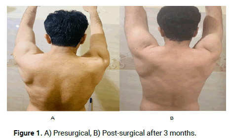
38-year-old orthopedic surgeon who noted right shoulder pain with shoulder movement in immediate postoperative period of lymph node resection in the right posterior triangle of neck (three weeks before clinic evaluation) as part of work up for an enlarged lymph node in neck. The pain was intermittent and dull in nature without any radiation in the arm. It occurred mainly on abduction of the right arm and shrugging of the shoulder with tenderness. His wife noticed atrophy of right shoulder and neck muscles. There was no relevant history of numbness, urinary incontinence, weight loss or weakness in the right upper extremity. He had no past medical or family history of note. On examination, there was atrophy of right trapezius muscle (upper part) and loss of bulk in right supra-scapular fossa. As the patient developed symptoms after surgery in posterior triangle, so there was concern for SAN injury during the surgical procedure. Nerve conduction study and electromyography was done with electrodiagnostic evidence of reduced Compound Muscle Action Potential (CMAP) almost 25% on right side compared to left spinal accessory nerve conduction study with mildly prolonged latency consistent with predominant axonal injury. There was electromyographic evidence of acute and chronic denervation changes in the right trapezius muscle in all parts. Due to recordable potentials after stimulating above the surgical scar, there was concern for partial right spinal accessory nerve dysfunction. Patients received surgical repair within 8 weeks of original insult and follow up nerve conduction studies showed more than 50% improvement in recording over trapezius muscle compared to initial study [6].
SAN injuries are difficult to diagnose however, timely diagnosis and management in experienced hands is important in such cases and can show promising results. Physical therapy shows marked improvement, however, there is a need for multidisciplinary management guidelines (Figure 1). Our case came to attention early due to the medical background of the patient followed by early diagnosis and surgical repair with near normal recovery (Tables 1-3).

Figure 1. A) Presurgical, B) Post-surgical after 3 months.
| Nerve/Sites | Muscle | Latency ms | Amplitude mV | Durationms | Rel Amp % | Segments | Distance cm | Lat Diff ms | Velocity m/s | Temp. °C |
|---|---|---|---|---|---|---|---|---|---|---|
| R Accessory (spinal)-Trapezius | ||||||||||
| Neck | Trapezius | 3.13 | 2 | 12.45 | 100 | |||||
| 2 | Trapezius | 3.54 | 1.7 | 12.6 | 87.2 | |||||
| 3 | Trapezius | 4.11 | 1.8 | 12.08 | 102 | |||||
| L Accessory (spinal)-Trapezius | ||||||||||
| Neck | Trapezius | 2.19 | 8.4 | 13.39 | 100 | |||||
| 2 | Trapezius | 2.34 | 5.5 | 12.34 | 66 | |||||
| 3 | Trapezius | 2.5 | 6.7 | 12.29 | 121 | |||||
Table 1. Pre-surgical Nerve conduction study.
| EMG summary table | |||||||||||
|---|---|---|---|---|---|---|---|---|---|---|---|
| Spontaneous | Recruitment | MUAP | |||||||||
| Muscle | Nerve | Roots | IA | Fib | PSW | Fasc | Others | Pattern | Amp | duration | PPP |
| R. Trapezius (lower) | Accessory (spinal) | C3-C4 | N | None | 1+ | None | Normal | N | Normal | Normal | N |
| R. Trapezius (upper) | Accessory (spinal) | C3-C4 | N | None | 3+ | None | Normal | N | Normal | Broad | 2+ |
| R. Trapezius (Middle) | C3-C4 | N | None | 2+ | None | Normal | N | Normal | Broad | N | |
Table 2. EMG summary table.
| Nerve / Sites | Muscle | Latency ms | Amplitude mV | Duration ms | Rel Amp % |
|---|---|---|---|---|---|
| R Accessory (spinal)-Trapezius | |||||
| Neck | Trapezius | 4.79 | 5.8 | 14.22 | 100 |
| 2 | Trapezius | 3.18 | 6.9 | 14.06 | 120 |
| 3 | Trapezius | 6.67 | 2.7 | 13.54 | 38.8 |
Table 3. Post-surgical nerve conduction study.
The knowledge about the functional and surgical anatomy as well as signs and symptoms produced after injury of the SAN is essential to avoid the injury and if it occurred, then early surgical management is necessary to prevent disability. Unfortunately, these lesions continue to occur and even though the symptoms may appear early, failure to recognize signs and symptoms results in delayed diagnosis even though the deficit is noticed early in some cases. Early referral for electrophysiological assessment can result in prompt surgical repair and can save loss of muscle function.
[Crossref] [Google Scholar] [PubMed]
[Google Scholar] [PubMed]
[Crossref] [Google Scholar] [PubMed]
[Crossref] [Google Scholar] [PubMed]
[Google Scholar] [PubMed]
[Crossref] [Google Scholar] [PubMed]
Citation: Khalid E, et al. "Iatrogenic Spinal Accessory Nerve Injury with Electrophysiological Assessment". Surg: Curr Res, 2023, 13 (11), 1-3.
Received: 10-Jul-2023, Manuscript No. SCR-23-25498; Editor assigned: 12-Jul-2023, Pre QC No. SCR-23-25498 (PQ); Reviewed: 26-Jul-2023, QC No. SCR-23-25498; Revised: 04-Oct-2023, Manuscript No. SCR-23-25498 (R); Published: 11-Oct-2023, DOI: 10.35248/2161-1076.23.13(10).456
Copyright: © 2023 Khalid E, et al. This is an open-access article distributed under the terms of the Creative Commons Attribution License, which permits unrestricted use, distribution, and reproduction in any medium, provided the original author and source are credited.