Mini Review - (2023) Volume 12, Issue 1
Gynecomastia is defined as benign glandular proliferation of breast tissue in males resulting in enlargement which can occur from hormonal imbalance caused by medications, foods, PIEDS, adolescence, obesity, narcotics, and even some fermented drinks. Historically there are various forms of grading and treatment depending on the patient’s presentation, including tanner staging, ultrasound, simon classification of gynecomastia, and concentric circles. All of these methods for grading are based on a physical exam, and treatment varies depending on if there is an underlying condition, which must be addressed first before pursuing surgery. Though there currently are some guidelines for management and diagnosis of gynecomastia, there is not a golden standard for diagnosis or surgery. The purpose of this study is to provide evidence to contribute towards setting a golden standard for diagnosis and treatment of gynecomastia through a new gynecomastia grading system with clearer guidelines.
Medications • Foods • Adolescence • Obesity • Narcotics
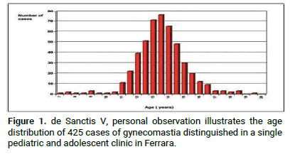
The rising prevalence of gynecomastia in men has significantly increased, affecting 35% of men, according to recent studies (Figure 1). It is the most prevalent benign disorder of the breast affecting men, typically between ages 50-69 [1-3].

Figure 1: de Sanctis V, personal observation illustrates the age distribution of 425 cases of gynecomastia distinguished in a single pediatric and adolescent clinic in Ferrara.
The distribution in the graph shows an increase in incidence from ages 11-17, this correlates with the onset of puberty and fluctuating hormone levels. New cases are emerging targeting newborns, adolescents and men over 50, in newborns the case is self-limiting.
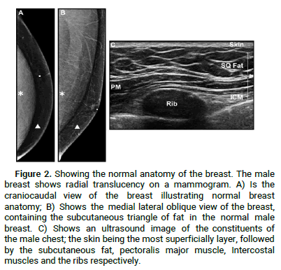
Speculation has driven the prevalence of gynecomastia, from the toxic hormone levels in the water negatively affecting fish, to higher levels of obesity leading to elevated oestrogen in men (Figures 2 and 3).
Anatomy of normal breast tissue (ultrasound)

Figure 2: Showing the normal anatomy of the breast. The male breast shows radial translucency on a mammogram. A) Is the craniocaudal view of the breast illustrating normal breast anatomy; B) Shows the medial lateral oblique view of the breast, containing the subcutaneous triangle of fat in the normal male breast. C) Shows an ultrasound image of the constituents of the male chest; the skin being the most superficially layer, followed by the subcutaneous fat, pectoralis major muscle, Intercostal muscles and the ribs respectively.
Assessment of gynecomastia

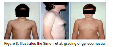
Figure 3: Illustrates the Simon, et al. grading of gynecomastia.
Simon, et al. classified 4 grades of gynecomastia, based on the size and outside appearance.
Grade I: A Small enlargement, with no excess skin.
Grade IIa: Moderate enlargement with no excess skin.
Grade IIb: Moderate enlargement with little excess skin.
Grade III: Marked enlargement with excess skin, imitating female breast ptosis.
Rohrich, et al. submitted a similar grading system of gynecomastia, similarly consisting of four grades.
Grade I: Minimal hypertrophy (<250 g) without ptosis.
Grade II: Moderate hypertrophy (250-500 g) without ptosis.
Grade III: Severe hypertrophy (>500 g) with grade I ptosis.
Grade IV: Severe hypertrophy with grade II or grade III ptosis.
Gland composition
Three main histological types of gynecomastia have been put forward to describe the composition of gynecomastia.
• Florid ductal proliferation and ductal hyperplasia characterizes
this classification of gynecomastia, edematous and loose
stroma is observed.
• Fibrous this type is characterized by stromal fibrosis and less
ducts.
• Intermediate this type is an intermediate between the two
previous types. Features of both Florid and Fibrous
gynecomastia are observed.
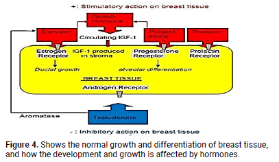
If the gynecomastia persist for longer than a year the fibrous type is more prevalent and becomes irreversible, which decreases the success of medical intervention (Figure 4) [4,5].
Hormonal imbalance

Figure 4: Shows the normal growth and differentiation of breast tissue, and how the development and growth is affected by hormones.
Estrogen, GH, IGF-1, progesterone and prolactin: The previously mentioned hormones have been shown to be integral parts of breast development.
Androgen and aromatase: These substances don’t directly stimulate breast development, they aromatize to estrogen which directly causes breast development.
Tumours: depending on the location and size of the tumour, estrogen may be released, which leads to breast development e.g. leading cell tumours, granulose cell tumours, adrenal tumour, sertoli cell tumours etc.
SHBG: Another cause of gynecomatia linked to estrogen incorporates steroid displacement from SHBG (Sex Hormone Binding Protein). Drugs such as spironolactone may cause this. This leads to displacement of oestrogen which allows more circulation of oestrogen in the blood.
Testosterone decrease and resistance to androgens: Androgens oppose the effect of estrogen, there is equilibrium between circulating levels of the two substances, which prevents males from developing male breast.
Decrease testosterone can also cause an imbalance in this equilibrium, leading to an elevation of estrogen circulating in the blood.
Thyrotoxicosis: Stimulation of the thyroid hormone on peripheral aromatase leads to elevated estrogen levels, thyrotoxicosis is strongly associated with gynecomastia.
Drugs: Approximately 20% of gynecomastia is caused by drugs or exogenous chemicals. The mechanism of action of these drugs differ, some may increase estrogen, while others possess estrogen like properties [6].
Recreational drugs such as marijuana, heroin, amphetamines and methadone have been linked with gynecomastia. Below is a Table 1 showing drugs that cause gynecomatia through known mechanisms.
| Mechanism | Drugs |
|---|---|
| Estrogen like, or blinds to estrogen receptor | Estrogen vaginal cream |
| Estrogen containing embalming cream | |
| Delousing power | |
| Digitalis | |
| Clomiphence | |
| Marjuana* | |
| Stimulate estrogen synthesis | Gandotrophins |
| Growth Hormones | |
| Supply aromatizable estrogen precursors | Exogenous androgen |
| Androgen precursors (i.e., androstenedione and DHE) | |
| Direct testicular damage | Busulfan |
| Nitrosurea | |
| Vincristine | |
| Ethanol | |
| Block testosteron sysnthesis | Ketaonazole |
| Spironolcatone | |
| Metronidazole | |
| Etomidate | |
| Block androgen action | Flutamide |
| Bicalutamide | |
| Finasteride | |
| Cyproterone | |
| Zanoterone | |
| Cimetidene | |
| Ranitidene* | |
| Spironolcatone | |
| Displace estrogen from SHBG | Spironolcatone |
| Ethanol | |
| *weak evidence | |
Table 1: Shows drugs that cause gynecomatia through known mechanisms.
While the previous grading systems have helped characterize and partly classify the level of gynecomastia into respective groups based on external phenotype, surgical experience has shown little is done to define the actual pseudo gland that is seen once operated on.
Gynecomastia pseudo glands are found to have an “iceberg appearance” where what is initially portrayed through visual interpretation and superficial examination varies significantly from what follows during surgical incision. Features such as tissue composition, shape and excess tailing portray significant impacts throughout the surgery and may lead to unforeseen complications.
The use of alternative imagining can help provide the necessary information to tackle such hurdles and prevent future issues from occurring.
An ultrasound scan will aid in differentiating the composition and thickness of the gland in addition to the border outline, this in turn will assist the surgeon and their surgical route. If the gynecomastia pseudo gland is classified in this manner, it will aid the surgeon to create a surgical plan and to foresee any upcoming surgical hurdles.
As a result of the many factors listed above, we are suggesting a change to this grading system. Our grading system dissects the original system in place further, while maintaining the original 1-4 grades but adding subsections. It is less of a superficial ranking, becoming more quantitative with regards to size while also categorizing the differences with tissue type, shape and tails further adding subsections accordingly.
The currently used and accepted grading system was put in place by Simon, et al. which is as follows:
Grade I: A Small enlargement, with no excess skin.
Grade IIa: Moderate enlargement with no excess skin.
Grade IIb: Moderate enlargement with little excess skin.
Grade III: Marked enlargement with excess skin, imitating female breast ptosis.
The system currently in use is qualitative which allows room for subjectivity which is not the best. The immense diversity in gynaecomastia simply is not recognized when this system is in use.
Skevofilax gynecomastia grading
Grade Ι: Overall small and not very noticeable. Ia and Ib may not need surgery and can be managed due to their flattened disc like shape. Ic and Id would likely require surgical intervention as these are more rounded and protruding.
• Ia=<5 cm, disc, no tails, adipose.
• Ib=<5 cm, disc, no tails, fibrous.
• Ic=<5 cm, sphere, no tails, adipose.
• Id=<5 cm, sphere, no tails, fibrous.
Grade II: Larger and more noticeable than grade I. All these subcategories would require surgical intervention. Tails start to show here (subnoted such that IIa (m) would be medial and IIa (l) would be lateral).
• IIa=6-10 cm, disc, adipose.
• IIb=6-10 cm, disc, fibrous.
• IIc=6-10 cm, sphere, adipose.
• IId=6-10 cm, sphere, fibrous.
Grade III: Largest in size but without excess dermal tissue. All require surgical intervention. Many have tails.
• IIIa=>10 cm, disc, adipose.
• IIIb=>10 cm, disc, fibrous.
• IIIc=>10 cm, sphere, adipose.
• IIId=>10 cm, sphere, fibrous.
Grade IV: All are large and have excess dermal tissue needing resection. Only two subcategories which are determined by the difference in adipose to fibrous. This makes a difference in the liposuction use.
• IIa=>10 cm, adipose, skin excess.
• IIb=>10 cm, fibrous, Skin excess.
The original systems put in place by Simon, et al. and Rodrich, et al. are in comparison not nearly as detailed. This proposed skevofilax gynaecomastia grading draws us closer to a golden standard which is so sought after in medicine, and can become a universal categorization method. These groupings have little to no overlap and allow for very little room for subjectivity.
Citation: Skevofilax IC, et al. "Does the Current Gynecomastia Grading System Accurately Correlate to Surgery". Reconstr Surg Anaplasto, 2023,12(1),1-4
Received: 14-Jul-2022, Manuscript No. ACR-22-18372; Editor assigned: 19-Jul-2022, Pre QC No. ACR-22-18372 (PQ); Reviewed: 02-Aug-2022, QC No. ACR-22-18372; Revised: 26-Dec-2022, Manuscript No. ACR-22-18372 (R); Published: 05-Jan-2023, DOI: 10.37532/acr.23.12.1.001-004
Copyright: © 2023 Skevofilax IC, et al. This is an open-access article distributed under the terms of the Creative Commons Attribution License, which permits unrestricted use, distribution, and reproduction in any medium, provided the original author and source are credited.