Research Article - (2023) Volume 8, Issue 1
Corona outbreak opens lots of scientific correlations of COVID virus with other similar virus. As it is highly mutating and contagious virus critical analysis and comparative studies are one of the most important tasks for identification of characteristic of this virus. Comparisons and co relations between the COVID-19 virus and influenza can be possible at different microscopic level. As COVID-19 outbreak continues to emerge and evolve, comparisons drawn as both cause respiratory disease, yet both have significant differences between them and how they spread. Computational examination is important and done by utilizing KEGG database for the influenza virus and tracing the equivalent for SARS-CoV-2, and furthermore by tabulating the biochemical, cellular and pathway contrasts between the two. It has important implications for public health measures and bioinformatics studies as the highly infective Coronavirus Disease-19 (COVID-19) is caused by a novel strain of Coronaviruses the Severe Acute Respiratory Syndrome Coronavirus-2 (SARS-CoV-2). This study becomes more imperative from the viewpoint to take all the necessary precautions and measures both on extrinsic front as public health and hygiene by following sanity measures as well as by following natural diet methods and plant based products which acts as immunity booster and all this will be possible only when we know the Scientific background for the same. Therefore, it is also important to generate awareness also.
COVID-19 • Influenza A virus • KEGG • Bioinformatics • Pathways • Corona virus
A virus on the basis of cell components predominantly and other parameters. The examination is important and done utilizing bioinformatics tool KEGG database and using disease database maps from KEGG for the influenza virus and tracing the equivalent for SARS-CoV-2 and furthermore by tabulating the biochemical, cellular and pathway contrasts between the two [1]. It has important implications for public health measures and bioinformatics studies as the highly infective Coronavirus Disease 19 (COVID-19) is caused by a novel strain of Coronaviruses the Severe Acute Respiratory Syndrome Coronavirus 2 (SARS-CoV-2) (Figure 1).

Figure 1: Transmission electron microscope image shows SARS-CoV-2 also known as 2019-CoV, the virus that causes COVID-19 isolated from a patient. The spikes on the outer edge of the virus particles give Coronaviruses their name, crown like.
A virus on the basis of cell components predominantly and other parameters. The examination is important and done utilizing bioinformatics tool-KEGG database and using disease database maps from KEGG for the influenza virus and tracing the equivalent for SARS-CoV-2 and furthermore by tabulating the biochemical, cellular and pathway contrasts between the two. It has important implications for public health measures and bioinformatics studies as the highly infective Coronavirus Disease 19 (COVID-19) is caused by a novel strain of Coronaviruses the Severe Acute Respiratory Syndrome Coronavirus 2 (SARS-CoV-2).
Influenza, also known as common flu is an acute respiratory illness which is highly contagious in nature. COVID-19 belongs to the same category of respiratory illnesses as influenza, but both are prompted by different viruses. New Coronavirus (known as SARS-CoV-2) is the major causative agent of COVID-19 and flu is the result of infection with influenza viruses [2,3].
The Corona Virus (CoVs), was first identified in late 1960’s, as a family of viruses that had the ability to infect wide range of hosts, such as humans, bats, rodents, civets, livestock and Arabian camels [4]. The impact of CoV infection on humans may range from gentle to severe depending on their genomic and phylogenetic features, thereby, altering the normal functions of respiratory system, Gastro Intestinal (GI) system and/or Central Nervous System (CNS) [5]. CoVs belongs to the Corovaviridae family of the viruses (Coronaviridae subfamily, nidovirales order), having single stranded, positive sense RNA as a genetic material, enclosed in a lipid bilayer. Amongst the four genera (α-, β-, γ- and δ-Coronavirus) of this virus, only α-Coronaviruses and β-Coronaviruses can infect the mammals.
In December 2019, a novel strain of Coronavirus the Severe Acute Respiratory Syndrome Coronavirus-2 (SARS-CoV-2) was discovered in the city of Wuhan, acting as an unknown pneumonia causing agent [6]. The origin of SARS-CoV-2 was credited to the zoonotic transfer of bat Coronaviruses, possibly through the animals. During early 2020, COVID-19 spread remarkably across all the continents and soon grew into a public health emergency. COVID-19 claimed millions of lives across the globe and ultimately was declared as a pandemic by the World Health Organization (WHO). The genome, protein structure and intracellular mechanisms of SARS-CoV-2 show an uncanny resemblance to the other members of the Coronavirus family that may transform into mild or asymptomatic to severe infectious conditions [7]. The underlying mechanism of the progression of this virus is still not clarified, although, many recent studies and evidences has suggested that SARS-CoV-2 may predominantly behave as other β- Coronavirus members. The key feature of this single stranded, positive sense RNA virus is the spike like glycoproteins.
It majorly constitutes the structural proteins like spike protein, envelope protein, membrane protein, and nucleo capsid protein. SARS-CoV-2 is a 30 kilobase genome, encoding viral proteins in almost 14 Open Reading Frames (ORFs) [8].
Fever and cough are the common symptoms associated with COVID-19, but other symptoms such as anosmia, cardiovascular and GI disorders have also been reported in many cases, thus, indicating the presence of multiple targets of the infection alongside the respiratory tract [9]. It has been observed that people with co-morbidities which are unrelated to the respiratory tract (such as hypertension, diabetes and cardiovascular disease) are at higher risk of getting infected with this virus. The primary target of the novel Coronavirus is the viral receptor, the Angiotensin Converting Enzyme (ACE) present on the human bronchial epithelium (Figure 2).

Figure 2: RNA genome is associated with the N protein to form the nucleocapsid.
The common flu or seasonal flu or influenza is caused by the infection of contagious influenza viruses that can affect the normal functioning of the respiratory system (throat, nose, and, sometimes, the lungs) and has the ability to cause seasonal, endemic infections in addition with periodic, unpredictable pandemics [10]. They belong to the family Orthomyxoviridaean, which are a group of antigenically and genetically diverse viruses. It is an enveloped negative strand RNA virus, containing seven to eight gene segments. Influenza a virus infects a wide variety of warm blooded animals, including birds, swine, horses, humans, and other mammals. Influenza virus replicates in the epithelial cell of both the upper and lower respiratory tract (Figure 3).

Figure 3: Structure of influenza virus.
NF-κB is a Nuclear Factor κB (NF-κB), is a transcriptional factor regulating the expression of more than 150 genes [11]. These genes are involved in governing the function of genes responsible for the production of cytokine/chemokines, cell adhesion and anti/pro-apoptotic genes. NF-κB is present as a complex with its inhibitor IκB. In the case of SARS-CoV-2 virus, the NFκB/IκB pathway plays a crucial role in mediating the inflammatory and immune responses against the infection, as well as, proliferation and apoptosis of the cells [12]. For the activation of this pathway, enzyme IκB Kinase (IKK) plays an essential role.
PI3K, belongs to the family of enzymes that play an important role in the proper functioning of the cell, cell survival, proliferation, differentiation, etc. The infection caused by influenza A virus, in turn, activates PI3K/Akt pathway, which acts as a critical checkpoint which regulates various cellular processes and signaling, like, growth, translation, vesicle trafficking, membrane composition and immunity [13]. Recent studies have suggested the role of viral NS1 protein in the efficient replication of the viral component.
In the current project paper we present the main aspects related to the SARS-CoV-2 virus and influenza a virus highlighting major differences as well as similarities and also the signaling and biochemical pathway contrast with the help of KEGG pathway.
Data collection
Together the information about the both, COVID-19 and influenza, various research articles, book chapters and newsletters were thoroughly studied and referred strain types, mode of transmission, symptoms, diagnosis and treatment [14]. The comparative study of the virus family, origin, and strain types, mode of transmission, symptoms, diagnosis and treatment was done for the better understanding of the similarities and dissimilarities between the two viruses. With the help of KEGG database, the map for influenza a virus was obtained.
KEGG: Kyoto Encyclopedia of Genes and Genome is a database that helps us to understand and analyze the high order functions and interactions of various units like cell, organism and ecosystem with each other [15]. To generate the output it takes into account the various large scale molecular datasets available which are generated using genome sequencing and other high-throughput technologies.
As COVID-19 is a recent disease turned pandemic, we have self-drawn a map for it, as we were unable to retrieve it from the KEGG database.
Why KEGG database?
Disease information is computerized in two forms: Pathway maps and gene/molecule lists. The KEGG pathway database contains pathway maps for the molecular systems. In the KEGG disease database, the representation of each disease is done in the form of list of known disease genes and much other underlying reason that may impact the molecular system such as environmental factors at the molecular level, diagnostic markers and therapeutic drugs [16].
PDB-RCSB
The structure of SARS-CoV-2 (PDB ID: 5I08) and influenza A virus was obtained from PDB.
Protein Data Bank (PDB) is a primary database and a comprehensive repository for the primary and secondary structure information of the proteins along with the coordinate’s data of the various atoms of the biomolecules (Figures 4 and 5).

Figure 4: PDB-RCSB Coronavirus structure and protein visualisation.

Figure 5: Home page of KEGG.
Flow chart
Go to Google→type KEGG and search→click on the KEGG website→KEGG home page opens→select KEGG pathway→go to pathway maps→choose infectious disease: Viral→ select influenza A viral disease→KEGG map for influenza A appears→take snapshot or download the map→paste on the document→trace the map accordingly for SARS-CoV-2 virus.
Comparison of pathway SARS-CoV-2 virus and influenza a virus (Figures 6 and 7).

Figure 6: KEGG disease pathway map for influenza A virus.

Figure 7: Disease pathway map-1 (self-drawn) for SARS-CoV-2 (COVID 19 virus).
The COVID-19 pandemic has made an adverse effect on the economic and public health infrastructure of many developed and developing countries across the globe. Understanding the pathway of the disease pathogenesis is critical for the diagnosis, prevention and the drug development [17]. The biochemical pathways of the SARS-CoV-2 virus and influenza A virus have been compared and analyzed in this paper.
Through the rigorous literature review, we have found some key similarities and dissimilarities between the two viruses. The self-drawn maps of the SARS-CoV-2 virus and the pathway map of the influenza a virus obtained from the KEGG database highlights the key surface proteins and genomic differences amongst the two. Influenza virus is a negative sense, single stranded and segmented RNA virus, having 4 strains and multiple subtypes. Influenza virus is an enveloped virus covered with Hemagglutinin (HA) and Neuraminidase (NA) proteins on its surface. On the other hand, COVID-19 virus has a positive sense and non-segmented single stranded RNA genome with only 1 strain. Unlike the influenza virus it has only spike protein(s) and no HA or NA surface proteins [18]. It is also an enveloped virus. The receptors for this virus are ACE2 proteins, present on the epithelial cells of lungs and small intestine.
The two main types of influenza virus type A and type B can cause mild to severe illness. The influenza type A has the ability to spread rapidly among the population may transform into pandemics and epidemics [19]. On the other hand, influenza type B is found only in humans and causes only mild illness (Table 1).
Table 1. Similarities and differences between COVID-19 virus and influenza a virus.
| Points of differences | COVID-19 virus | Influenza A |
|---|---|---|
| Family | Coronaviridae | Orthomyxoviridae |
| Strains | 1 strain | 4 strains, multiple subtypes |
| Mode of transmission | Person to person contact and through direct contact with respiratory droplets | Mainly by droplets made when people with flu cough, sneeze or talk |
| Symptoms | Fever, dry cough, tiredness are most common symptoms | Fever, chills, muscle aches, cough, congestion, runny nose, headaches and fatigue. |
| Incubation period | 1 to 12.5 days | 1 to 4 days |
| Reproductive number | 2 to 2.5 | 1.13 |
| Genome | (+) Strand non segmented RNA genome | (-) Strand single stranded segmented RNA genome |
| Surface proteins | Four main structural proteins contain Spike (S), Membrane (M), Envelope (E) and Nucleocapsid (N) Proteins | Hemagglutinin and Neuraminidase |
| Protein functions | Spike protein-(S) binding of host cell receptors to facilitate entry of host cell; Nucleocapsid protein-(N) Bound to RNA genome to make up nucleocapsid; Envelope protein-(E) Form viral envelope; Membrane protein-(M) Central organiser of CoV assembly | Hemagglutinin-(H) needed for infection; its presence inhibits release of the particle after budding. Neuraminidase-(N) cleaves terminal sialic acid residues from glycan structures on the surface of the infected cell. |
| Diagnosis | Viral tests and antibody tests | Viral culture, serology, rapid antigen testing |
| Treatment | No specific drugs are available till now. Few precautionary vaccines are available like Covishield, Covaxin, Pfizer | Relenza and Tamiflu. Antiviral vaccines, supportive and self-care and medication are there |
| Vaccine availability | No active vaccine available | Seasonal vaccine and antiviral medication available |
| Size | 26 and 32 kB | 13.5 kb |
| Pathway | NF-κB pathway | PI3K-Akt pathway |
| Vulnerable groups | Older age people | Children, elderly and women |
The early symptoms of COVID-19 may include fever, tiredness, dry cough, red eyes, and in some cases, loss of taste, and smell. Some patients may experience nasal congestion, sore throat, and diarrhea. As the disease progresses inside the body, symptoms may worsen and the patient might experience shortness of breath or difficulty breathing.
The in depth study of various resources allowed us to conclude that both, the influenza and COVID-19 possesses many common symptoms which may eventually lead to pneumonia. While the symptoms are similar in both diseases, the fraction with severe disease is higher for COVID-19.
Also, through literature studies we established the fact that both the viruses vary significantly in their pathways, proteins, receptors, reproductive strength and genomic composition. SARS-CoV-2 follows NF-κB pathway and influenza A virus utilizes PI3K–Akt pathway. Comparative study of both the viruses has been tabulated on the basis of their pathways maps. We all are very well aware of the fact that prevention is better than cure. While there is no medicine for COVID-19 as of now, taking preventive measures and working on our immunity with special emphasis on our respiratory health is our ultimate weapon of combat against this deadly virus. There are many ancient Ayurveda literatures and scientific literatures that propose simple and effective remedies to improve and maintain our body’s immune response against such viruses. Table 2 consists of few medicinal plants along with their active compounds and structures which can potentially boost our immunity.
Table 2. Immunity boosting plants against COVID-19 Pandemic with their active constituents and structures.
| S. No. | Botanical name | Common name | Family | Active constituent | Structure of active constituent |
|---|---|---|---|---|---|
| 1 | Curcuma longa | Turmeric | Zingiberaceae | Curcumin (diferuloylmethane) |
|
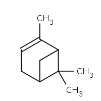
| 2 | Cuminum cyminum | Cumin | Apiaceae | Cuminaldehyde and Pinene |
|
| 3 | Coriandrum sativum | Coriander | Apiaceae | Linalool and Limonene |
|
| 4 | Allium sativum | Garlic | Amaryllidaceae | Diallyl thiosulfonate (allicin) |
|
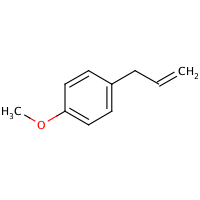
| 5 | Ocimum basilicum | Basil | Lamiaceae | Linalool and Methyl Chavicol |
|
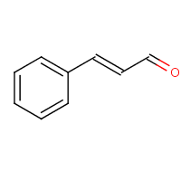
| 6 | Cinnamomum verum | Cinnamon | Lauraceae | Cinnamaldehyde and Cinnamate |
|
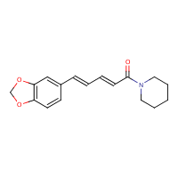
| 7 | Piper nigrum | Black pepper | Piperaceae | Piperine |
|
| 8 | Zingiber officinale | Ginger | Zingiberaceae | Zingerone and Gingerols |
|
| 9 | Mentha | Mint | Lamiaceae | Menthone |
|
| 10 | Syzygium aromaticum | Clove | Myrtaceae | Beta-caryophyllene and Eugenol |
|
Data were analyzed on GraphPad Prism software. Statistical significance of differences among cytokine levels was assessed using the Mann–Whitney U-test for nonparametric data. Associations between cytokine levels and hospitalization time (in days) were tested using Spearman rank-order correlation coefficient and visualized using the corrplot R package. Statistical significance of differences among gene groups was assessed using a two-tailed Student’s t-test for parametric data. Polar charts from the ggplot2 R package were used for the visualization of differences in cytokine response patterns.
The authors declare no conflict of interest for the research paper.
We would like to express our special thanks of gratitude to Dr. Anamika Singh and respected principal, Maitreyi college, university of Delhi for their support and encouragement. Who gave the golden opportunity to write the research paper on the topic "using KEGG pathways comparing the cell components of coronavirus and influenza virus".
[Crossref] [Google Scholar] [PubMed]
[Crossref] [Google Scholar] [PubMed]
[Crossref] [Google Scholar] [PubMed]
[Crossref] [Google Scholar] [PubMed]
[Google Scholar] [PubMed]
[Crossref] [Google Scholar] [PubMed]
[Crossref] [Google Scholar] [PubMed]
[Crossref] [Google Scholar] [PubMed]
[Crossref] [Google Scholar] [PubMed]
[Crossref] [Google Scholar] [PubMed]
[Crossref] [Google Scholar] [PubMed]
[Crossref] [Google Scholar] [PubMed]
[Crossref] [Google Scholar] [PubMed]
[Crossref] [Google Scholar] [PubMed]
[Crossref] [Google Scholar] [PubMed]
[Crossref] [Google Scholar] [PubMed]
[Crossref] [Google Scholar] [PubMed]
Citation: Singh A. "Comparative Pathway Analysis of Coronavirus and Influenza Virus". J Forensic Path, 2023, 8(1), 1-6.
Received: 26-Dec-2022, Manuscript No. JFP-22-21170 ; Editor assigned: 28-Dec-2022, Pre QC No. JFP-22-21170 (PQ); Reviewed: 11-Jan-2023, QC No. JFP-22-21170 ; Revised: 17-Mar-2023, Manuscript No. JFP-22-21170 (R); Published: 27-Mar-2023, DOI: 10.35248/2684-1312.23.8.1.350
Copyright: © 2023 Singh A, This is an open-access article distributed under the terms of the Creative Commons Attribution License, which permits unrestricted use, distribution, and reproduction in any medium, provided the original author and source are credited.