Case Series - (2022) Volume 11, Issue 5
The author reports on two cases of patients injecting liquid silicone into the mammary gland parenchyma, one patient injected silicone more than twenty years ago, and had many surgeries but did not remove all silicone, and the second patient was the first time. come to our hospital she wants to get silicone injected more than ten years ago. Both patients had palpable lumps on both sides of the mammary gland, there were no clinical changes in the skin where silicone lumps were palpable. Current ultrasound and magnetic resonance imaging images also show free silicon infiltration in the bilateral mammary gland tissue, concurrently infiltrating the pectoral muscles and reactive inflammatory lymph nodes in the armpit. The current treatment strategy is the surgical removal of infiltrated silicone in breast tissue combined with immediate breast implant placement to create aesthetics for the patient.
Liquid silicone • Autologous breast augmentation • Breast implant placement
Breast augmentation surgery is one of the top five most-performed cosmetic surgeries in the world [1]. Surgeons can choose from breast augmentation with breast implants, autologous breast augmentation with muscle flaps, or injections of fillers such as autologous fat or synthetic biomaterials (Paraffin, Silicone, PAAG [Polyacrylamide] hydrogel) [2]. Of these, liquid silicone has been used for soft tissue injections for the past six decades. In the early days of its development, silicone was known to be chemically inert, non-carcinogenic, readily shaped to soft tissues, and non-proliferative of bacteria [3]. Injecting silicone directly into the soft tissue of the mammary gland for reconstruction is currently banned, but complications of liquid silicone injection in the past years are still commonly reported in the literature such as infection, sensitization, and embolism. Vascular occlusion, pulmonary embolism, granulomatous reactions, nodules, skin discoloration, silicone infiltration in tissues [4,5], and recent reports of breast cancer reveal associated with silicone injections liquid [6-9].
Liquid silicone can be surgically removed and the mammary gland can be reconstructed with autologous breast augmentation or implant placement in a single surgery. In this report, the author reports 2 women who had undergone liquid silicone injections, of which 1 patient had undergone previous silicone removal surgery and one patient had undergone silicone removal surgery for the first time. We used the inverted T-line for the patient who had a previous surgical scar, and the second patient through the semi-areolar line to increase the dissection efficiency and access the mammary gland parenchyma for the patient. After removing the liquid silicone, both patients received immediate breast implants.
Case I
f multiple painful lumps in both breasts. The patient used to have liquid silicone injected into the mammary gland twenty-two years ago, in the past six years; the patient has undergone three surgeries at other hospitals to remove the silicone. When coming to the Department of Plastic SurgeryEmcas Plastic Surgery Hospital, through clinical examination, we found lumps on both sides of the patient's mammary glands, scattered from the areola to the external mammary gland tissue. The lumps size ranges from a few millimeters to centimeters, non-motile; no change in skin color has been observed (Figure 1). Through external examination, the patient's silicone infiltration was classified as class II according to the classification of Ueno 1978 and not cause changes in the external appearance of the mammary gland [10]. Ultrasonography was limited to investigation in this patient due to the acoustic opacity of the free silicone structures. The patient’s breast Magnetic Resonance Imaging (MRI) was 3.0 Tesla, which recorded abnormal signal nodules, high signal on Short-TI Inversion Recovery (STIR) silicone-saturated sequences, and low signal on STIR silicone-saturated sequences suitable for silicone properties. The nodules were distributed in different sizes, scattered in the two mammary glands, with infiltrates into the pectoral muscles and reactive inflammation in the axillary nodes (Figures 2 and 3).
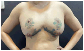
Figure 1: Patient 1 â?? Preoperative picture
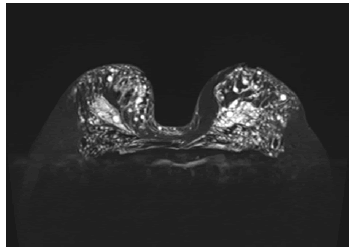
Figure 2: Magnetic resonance image of the patient's mammary gland before surgery.
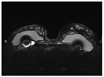
Figure 3: Magnetic resonance imaging of the patient's mammary gland after surgery.
Combining the history, laboratory tests, imaging, and in the patient's history with lumps and chronic pain, breast cancer screening at present has not found the patient can develop cancer. We advise patients to perform plastic surgery with autologous breast implants: Transverse Rectus Abdominis Myocutaneous Flap (TRAM), but if the patient wants to limit surgical areas on the body and soon recovered, the patient decided to choose breast implants. We performed a double mastectomy with siliconeinfiltrated breast tissue, the surgical incision was chosen as Inverted-T because the patient had undergone surgery in this way. We kept some of the blood vessels in the upper internal mammary perforator to protect the bilateral nipples, combined with immediate cosmetic reconstruction for the patient by placing the breast implant inside the space under the pectoralis major. However, during the surgery, we encountered a lot of liquid silicone particles causing local inflammatory reactions; the structure of the mammary gland tissue can be seen in color and stiffer than the surrounding tissues (Figure 4). The rupture of the silicone granules during surgery reduces the surgeon's ability to thoroughly dissect, especially in areas of silicone infiltration into the pectoralis major. We performed maximal dissection of free liquid silicone and minimally invasive to preserve the perfusion of the upper skin tissue, the amount of silicone and mammary tissue dissected on both sides were similar to each other (Figure 5). The histopathological results of the mammary gland parenchyma showed the proliferation of macrophages, lymphocytes, and neutrophils in the free silicone, with no malignant changes on the microscopic level (Figure 6).
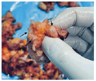
Figure 4: Infiltrated liquid silicone vacuoles in mammary gland tissue (Black arrows).
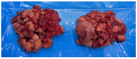
Figure 5: Amount of silicone infiltrating mammary gland tissue removed.
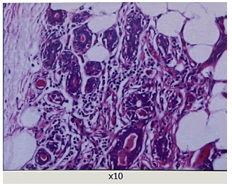
Figure 6: Histopathological results of the first patient with H&E staining with x10-objective.
After the surgery, the patient was monitored at the hospital for three days and then discharged, the patient's condition was stable, there was no more pain in the mammary glands and no silicone lumps were palpable under the skin. After three weeks, the patient returned and we checked the breast MRI image to assess that the amount of silicone remaining compared to the baseline had decreased much (Figures 3 and 7).
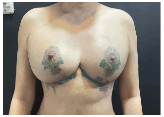
Figure 7: Picture of the first patient 1 week after surgery
Case II
A thirty-three-year-old female patient came to our hospital because of lumps in her mammary gland (Figure 8). The patient received a liquid silicone injection twelve years ago; the amount of liquid silicone injected into each side was about 200 cc, performed at a beauty salon abroad with the advertisement that it was "natural collagen". Through examination, we also noted lumps ranging in size from a few millimeters to centimeters, scattered on both sides of the mammary gland, painless, non-motile, and did not cause changes in skin color. Similarly, through clinical examination, we classified the patient as grade II according to Ueno 1978 [10]. The patient underwent imaging studies such as ultrasound and MRI, and the risk assessment for cancer was not noted. Ultrasound imaging is limited due to the scattered distribution of liquid silicone. The MRI results showed abnormal signal nodules, the largest size was 10 mm x 14 mm in the breast (T) and 11 mm x 12 mm in the breast (P), clearly demarcated (Figures 9 and 10). The patient has inflammatory lymph nodes in the armpit area; the size of the lymph nodes is about 10 mm x 5 mm, which is thought to be reactive inflammation.
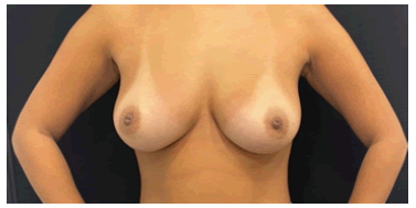
Figure 8: Second patient - preoperative picture.
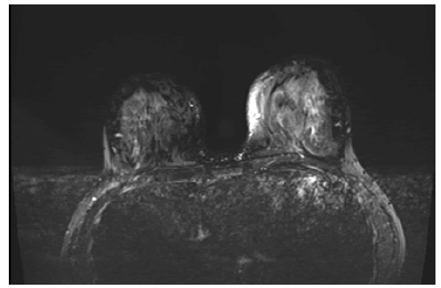
Figure 9: Magnetic Resonance Imaging (MRI) on STIR silicone-saturated sequences.
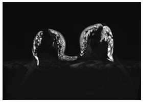
Figure 10: Magnetic resonance imaging (MRI) on STIR silicone-selective sequences.
As this is the first time silicone removal surgery, it is better to reconstruct the mammary gland after removing the patient's silicone with autologous breast augmentation (TRAM flap), which reduces the use of artificial biomaterials. However, the patient was young and wanted to have children in the future, so the patient chose to have a breast implant to preserve the integrity of the abdominal muscles and expect the earliest recovery results. Unlike the first case, we selected the surgical incision on the upper areola line, which minimized scarring and eased access to the entire mammary gland. We also encountered silicone nodules which break down into a liquid form, causing mammary parenchymal cutting and silicone removal to be hindered and possibly missed (Figures 11 and 12). After surgery, histopathology showed amorphous ingestion of macrophages, lymphocytes, and neutrophils (Figure 13). After the surgery, the patient was stable and continued to stay under observation at our hospital for two days, and then the patient was discharged. At present, the patient has no palpable breast lumps (Figure 14).
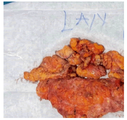
Figure 11: . Liquid silicone nodules
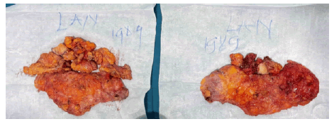
Figure 12: The amount of silicone-infiltrated mammary gland parenchyma removed.
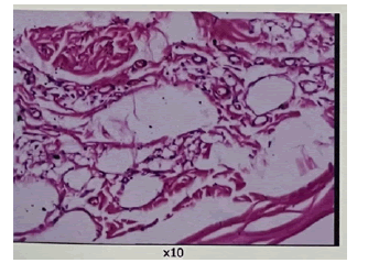
Figure 13: Anatomy results of the second patient with H&E staining with x10- objective.
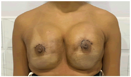
Figure 14:Picture of the second patient two days after surgery
Silicone was first isolated in 1824 by the Swedish chemist Johann Berzelius; however, he was primarily interested in the purity of silicone. The use of liquid silicone for injection into soft tissues became widespread worldwide in the 1940s [11], proceeded by the popularity of Paraffin (1899- 1914) and other biomaterials (1914-1943) [2]. Originally silicone was used in its pure form, but other substances such as vegetable oils were later added to increase local tissue reactivity and reduce migration, especially when applied with mass injections of silicone [12]. Complications of liquid silicone injections have been reported more frequently and the FDA has declared them illegal since 1970, and since 1991 silicone has been listed as prohibited for medical use except for certain surgical procedures [11, 13]. In Vietnam, the Ministry of Health has banned the injection of liquid silicone directly into soft tissues since 1995. However, in practice, liquid silicone is still used irregularly, in improving the aesthetic contours of patient’s nose, as well as acne scars, at the lowest cost without undergoing surgery [14,15].
Complications associated with liquid silicone injections have been reported to range from mild to severe, occurring 8 years-10 years after silicone injection (may persist for as long as 6 years-36 years) [11]. Minor complications include injection site reactions such as pain, erythema, ecchymosis, and edema [11, 16]. Serious complications such as granuloma formation, silicone displacement, nodules, cystic lesions, chronic cellulitis, pneumonia, embolism, cancer-associated disease, and death have been reported [2, 4, 6-8, 16-20].
In both of our patients, there was the infiltration of injected liquid silicone; assessment by magnetic resonance MRI is a useful method at this time to assess the extent of infiltration and to quickly facilitate the treatment, ability to differentiate from other breast tumors, and evaluate for metastasis [21, 22]. Combining clinical examination, laboratory test, imaging, and pathological results of mammary gland tissue after resection, we have not seen the possibility of malignancy in this patient. . In a study of 696 patients, Ohtake reported that 4.2% of patients had cancer associated with injections of liquid silicone and breast fillers, however, there was no clear evidence that silicone injections increased risk of cancer in breast tissue in women in women [23]. Similarly, Uchida et al reported that out of 3930 breast cancer surgeries, only 8 (0.2%) of breast cancers were associated with breast implants or artificial biomaterials [9]. However, changes in mammary parenchyma induced by liquid silicone injections make it difficult to interpret clinical signs and mammograms and thus may reduce the early diagnosis of breast cancer [21].
When liquid silicone persists in the body for a long time, it will stimulate inflammatory and fibrogenic processes [15], the mammary gland tissue is benign and the outer skin of the mammary gland has not been deformed, we classify it as grade II according to Ueno and et al. 1978, the decision to proceed to surgery on both sides of this patient was necessary before the silicone fraction could cause other changes in the patient [10]. In a case series of 28 patients (mean age 37 years) with moderate complications after 9 years of breast augmentation with liquid silicone injection, Parson et al recommended surgical removal of the breast. The mammary gland parenchyma was removed to preserve the nipple, but in some cases of small, uncomplicated liquid silicone, the authors agreed that only full follow-up was required, not necessarily surgery [24]. In our two patients, although case 1 had undergone surgical removal of silicone many times before, when performing surgery on both patients, the omission may appear due to the migration and infiltration of silicone into other tissues. Therefore, evaluation was necessary by MRI after surgery, to get the necessary follow-up treatment for the patient.
We chose the areola line for the first time silicone removal, to have easy access to the entire mammary gland parenchyma as well as the infiltrated silicone in the parenchyma. From 2003 to 2018, Bei Qian et al., when performing surgery on 325 patients, selected the areola line to maximize the effectiveness in removing PAAG as well as infiltrated breast parenchyma [25]. In another study, Megumi, after surgical removal of the infiltrated liquid silicone in the mammary gland, performed axillary breast implant surgery on 14 patients; however, 7 of them had complications of early necrosis remaining breast tissue, requiring the removal of breast implants and management of complications [26]. Therefore, the choice of an incision with easy access to the silicone site in the infiltrated mammary gland tissue, and at the same time avoiding complications after surgery, needs to be considered for each patient. Delayed cosmetic reconstruction of the breast after a mastectomy due to concerns about bleeding, however, if preserved tissue viability is satisfactory, external cosmetic reconstruction mammography can be performed within 1 week of mastectomy [24]. Kim et al. reported two early mastectomy surgeries with filler injected into the mammary gland, the authors recommended the use of autologous fat or the use of autologous muscle flaps, however depending on the amount of pectoral muscle and mammary gland tissue remaining after surgery, surgeons consider the choice of breast reconstruction method for patients [27]. For our patients, to reduce the risk of the patient having to undergo multiple surgeries, we choose to have the breast implant placed immediately after silicone removal to reduce the patient's need to undergo multiple surgeries and early recovery.
Citation: Khiem, P. X, et al. Case Report: Surgical Removal of Liquid Silicone Injected into the Mammary gland and Immediate Placement of Breast Implants on Perennial Liquid Silicone Injection Patient. Reconstr Surg Anaplastol, 2022, 11(5), 003-006
Received: 18-Oct-2022, Manuscript No. ACR-22-20411; Editor assigned: 25-Oct-2022, Pre QC No. ACR-22-20411 (PQ); Reviewed: 29-Oct-2022, QC No. ACR-22-20411 (Q); Revised: 05-Nov-2022, Manuscript No. ACR-22-20411 (R); Published: 10-Nov-2022, DOI: DOI.10.37532/22.11.5.003-006
Copyright: ©2022 Khiem, P. X, et al, This is an open-access article distributed under the terms of the Creative Commons Attribution License, which permits unrestricted use, distribution, and reproduction in any medium, provided the original author and source are credited.